Portable Ultrasound Scanner XF300 LED
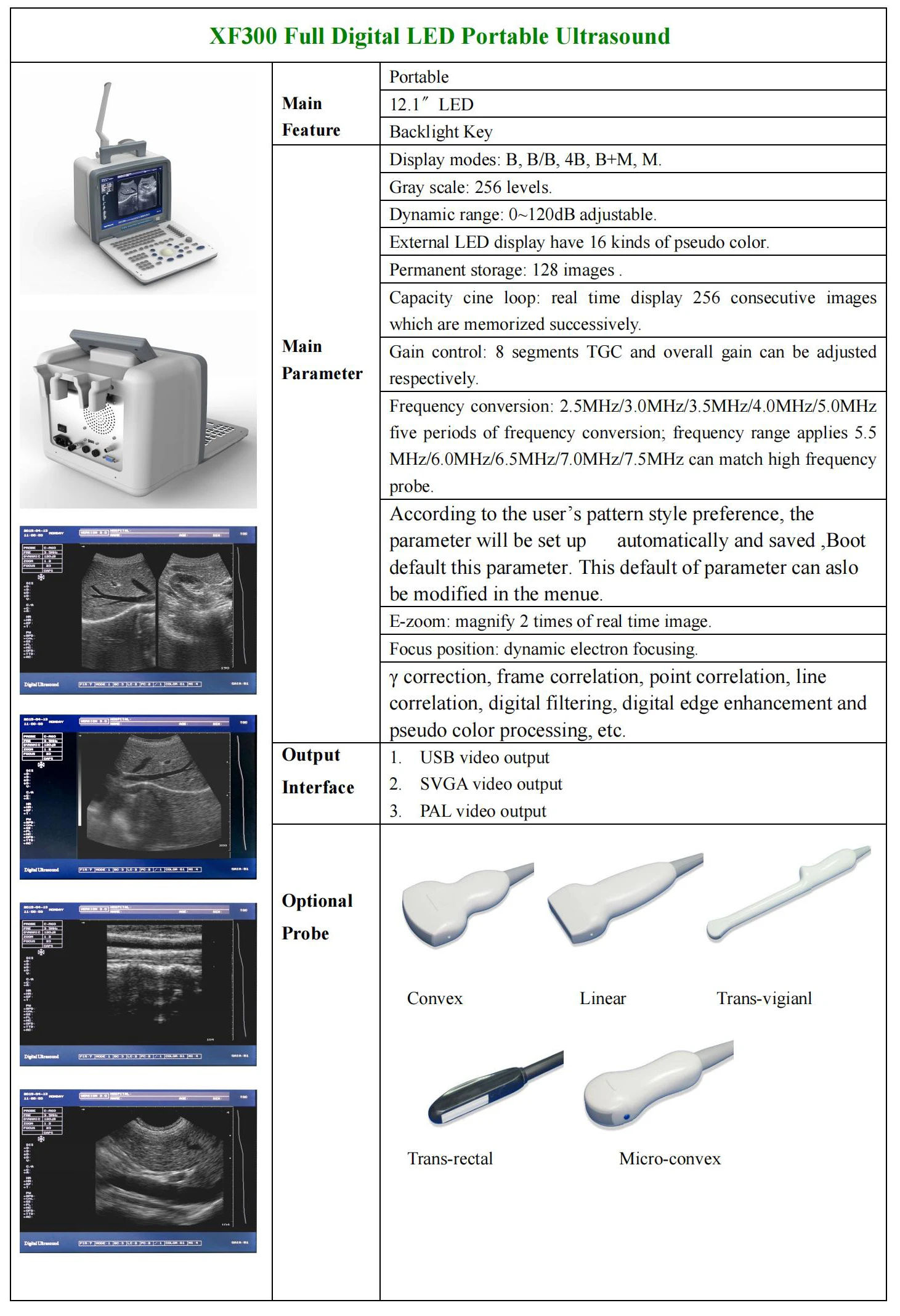
XF300 Full Digital LED Portable Ultrasound
How do ultrasound scans work?
An ultrasound scan uses high-frequency sound waves to create images of the inside of the body. It is suitable for use during pregnancy.
Ultrasound scans, or sonography, are safe because they use sound waves or echoes to make an image, instead of radiations
Ultrasound scans are used to evaluate fetal development, and they can detect problems in the liver, heart, kidney, or abdomen.
They may also assist in performing certain types of biopsy. The image produced is called a sonogram.
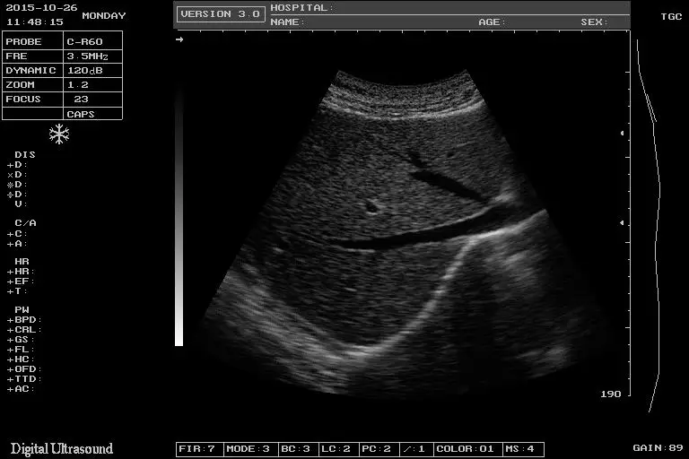
How does it capture an image?
Ultrasound will travel through blood in the heart chamber, for example, but if it hits a heart valve, it will echo, or bounce back.
It will travel straight through the gallbladder if there are no gallstones, but if there are stones, it will bounce back from them.
The denser the object the ultrasound hits, the more of the ultrasound bounces back.
This bouncing back, or echo, gives the ultrasound image its features. Varying shades of gray reflect different densities
XF300 LED Portable Ultrasound Scanner
Display Modes: 3.5MHz/R60 convex array probe:B,B/B,4B,B+M,M
5 MHz/R20 Dimpling array probe:B
6.5MHz/R13 Dimpling array probe:B
7.5 MHz/L40 Line array probe:B
Image Mutiplying Factor:
3.5MHz/R60 convex array probe: x0.8, x1.0, x1.2, x1.5,x1 .8, x2.0 ( 6modes)x0.8, x2.0 (display penetration depth)
6.5MHz/R13 Dimpling array probe: x0.8, x1.0, x1.2, x1.5 (4 modes)
7.5MHz/L40 Line array probe: x0.8, x1.0, x1.2, x1.5 (4 modes)
E-zoom:magnify 2 times of real time image
Dynamic Range:0~120dB ajustable
Focus Position:1,2,3 and 4-segments dynamic electron focusing
Image Processing: γcorrection, frame correlation,point correlation, line correlation, digital filtering, digital edge enhancement and pseudo color processing, etc.According to the user’s pattern style preference, the parameter will be set up automatically and saved ,Boot default this
parameter. This default of parameter can aslo be modified in the menue.
Frequency Conversion:2.5MHz/3.0MHz/3.5MHz/4.0MHz/5.0MHz five periods of frequency conversion
frequency range applies 5.5 MHz/6.0 MHz/6.5 MHz/7.0 MHz/7.5MHz can match high frequency probe
Measuring Function:Distance,circumference/area (method of ellipse, method of loci), volume, heart rate, gestational weeks (BPD,GS,CRL,FL,HL,OFD,TTD,AC), expected date of confinement and fetus weight, etc.
Annotation Function:hospital name, patient’s name, age and gender 64 body marks (with probe’s position);Full-screen character annotation; Real-time clock display
Puncture Guide:3.5MHz convex array probe can display puncture guide line in B mode
Gain Control:8 segments TGC and overall gain can be adjusted respectively
Image Polarity:left and right flip, black and white flip, up and down flip
Capacity Cine Loop: real time display 256 consecutive images which are memorized successively
Image Playback:series playback or check one by one
Permanent Storage:128 images
Output Interface:VGA video output offers connection to SVGA color display PAL video output offers connection to PAL display and printer video image recorder and image workstation, etc
USB Port:Offers storing images in USB flash disk
Features:
1. Mass memory , cine loop and permanent storage.
2. Radium Caved buttons and Silicon backlit buttons to be chosen;
3. Intelligent fluorescence trackball.
4. 8-segment TGC control. Overall Gain control
5. 2 USB ports , Upgraded and expandable printer function;
6. 2probe connectors. Probe automatic identfication;
7. 2SVGA video outputs and 21 PAL video outputs offer connection to video image recorder and image workstation. etc.
8. Report page is generated automatically.
9. According to the user's pattern style preference, the parameter will be set up automatically and saved ,Boot default this
parameter. This default of parameter can aslo be modified in the menue.
XF300 Full Digital LED Portable Ultrasound
| Model | Portable Led Ultrasound Scanner Machine XF300 S300 |
| Display Modes | 3.5MHz/R60 convex array probe:B,B/B,4B,B+M,M 5 MHz/R20 Dimpling array probe:B 6.5MHz/R13 Dimpling array probe:B 7.5 MHz/L40 Line array probe:B |
| Image Mutiplying Factor | 3.5MHz/R60 convex array probe: x0.8, x1.0, x1.2, x1.5,x1 .8, x2.0 ( 6modes)x0.8, x2.0 (display penetration depth) 6.5MHz/R13 Dimpling array probe: x0.8, x1.0, x1.2, x1.5 (4 modes) 7.5MHz/L40 Line array probe: x0.8, x1.0, x1.2, x1.5 (4 modes) |
| E-zoom | magnify 2 times of real time image |
| Dynamic Range | 0~120dB ajustable |
| Focus Position | 1,2,3 and 4-segments dynamic electron focusing |
Image Processing: | γcorrection, frame correlation,point correlation, line correlation, digital filtering, digital edge enhancement and pseudo color processing, etc. |
| According to the user’s pattern style preference, the parameter will be set up automatically and saved ,Boot default this parameter. This default of parameter can aslo be modified in the menue. | |
| Frequency Conversion | 2.5MHz/3.0MHz/3.5MHz/4.0MHz/5.0MHz five periods of frequency conversion frequency range applies 5.5 MHz/6.0 MHz/6.5 MHz/7.0 MHz/7.5MHz can match high frequency probe |
| Measuring Function | Distance,circumference/area (method of ellipse, method of loci), volume, heart rate, gestational weeks (BPD,GS,CRL,FL,HL,OFD,TTD,AC), expected date of confinement and fetus weight, etc. |
Annotation Function
| hospital name, patient’s name, age and gender 64 body marks (with probe’s position); Full-screen character annotation; Real-time clock display |
Puncture Guide | 3.5MHz convex array probe can display puncture guide line in B mode |
| Gain Control | 8 segments TGC and overall gain can be adjusted respectively |
| Image Polarity | left and right flip, black and white flip, up and down flip |
| Capacity Cine Loop | real time display 256 consecutive images which are memorized successively |
Output Interface | VGA video output offers connection to SVGA color display PAL video output offers connection to PAL display and printer video image recorder and image workstation, etc |
| USB Port | Offers storing images in USB flash disk |